
Imaging, QoL and Patient-Reported Outcome and Supportive Care
Category: Imaging, QoL and Patient-Reported Outcome and Supportive Care
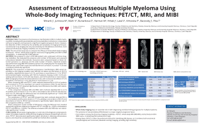
Assessment of Extraosseous Multiple Myeloma Using Whole-Body Imaging Techniques: PET/CT, MRI, and MIBI
 photo")
Jiří Minařík, MD, PhD (he/him/his)
Department of Hemato-Oncology
University Hospital Olomouc
The proportion of detected extraosseous manifestations (EM) varied by imaging modality. MRI detected no EM lesions in 92.5% of patients, one lesion in 6.3%, and three or more in 1.3%. PET/CT was more sensitive: 78.8% had no EM, 13.8% had one, 3.8% had two, and 3.8% had three or more lesions. MIBI scintigraphy had the lowest detection: 97.5% showed no lesions, 1.3% one lesion, and 1.3% three or more.
Comparing MRI and PET/CT, both detected EM in 8.8% and no lesions in 77.5% of patients, with an overall concordance of 86.3% and discordance in 13.7%. PET/CT revealed significantly more EM than MRI (p = 0.001).
MRI vs. MIBI showed EM detection in 2.5% and no lesions in 91.3%, with 93.8% concordance and 6.3% discordance. The difference was not statistically significant (p = 0.063), though MRI had slightly better sensitivity.
PET/CT vs. MIBI: both detected EM in 2.5% and no lesions in 77.5%, with 80.0% concordance and 20.0% discordance. PET/CT showed a significantly higher EM detection rate (p < 0.0001).
Wilcoxon test showed significant differences in lesion numbers between PET/CT and MRI (p = 0.003), and PET/CT and MIBI (p = 0.0004); the MRI vs. MIBI difference was not significant (p = 0.076).
Conclusions: Whole-body imaging plays a crucial role in the diagnosis and prognostication of multiple myeloma. While all three imaging techniques showed high concordance, PET/CT demonstrated significantly higher sensitivity in detecting extraosseous lesions and should be considered the preferred method when available.
Supported by MH CZ – DRO (FNOl, 00098892) and IGA_LF_2025_005