
Myeloma Novel Drug Targets and agents
Category: Myeloma Novel Drug Targets and agents
Secondary clones identified by mass spectrometry in baseline samples remain stable or disappear early during monitoring in multiple myeloma patients

Jill Pauli, PhD
Associate Director, Scientific Affairs
The Binding Site, part of Thermo Fisher Scientific
Mass spectrometry offers improved sensitivity over electrophoretic methods for identifying and quantifying monoclonal proteins. Besides the main M-protein, it can identify secondary, low-level M-proteins not previously reported by standard methods, with unknown clinical significance. We report on dynamic changes of secondary M-proteins identified by mass spectrometry and their clinical relevance in patients with active disease.
Methods:
The study included 62 intact immunoglobulin multiple myeloma patients, with at least one follow-up sample taken within 6(±3) months from baseline. Serum samples were analyzed with the Immunoglobulin Isotypes (GAM) for the EXENT® Analyzer (The Binding Site, part of Thermo Fisher Scientific). M-proteins were identified and quantified in baseline samples by the EXENT System and compared to immunofixation and serum protein electrophoresis (SPE) results. Secondary M-proteins were defined as monoclonal intact immunoglobulins (IgG, IgA, IgM) < 0.200 g/L and ≥0.015 g/L, or kappa and lambda only peaks with no associated heavy chain. Persistent M-proteins were defined by their presence during follow-up, based on the same m/z value (±4).
Results:
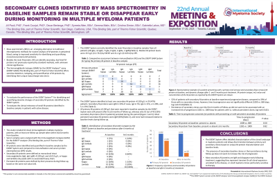
The EXENT System correctly identified the main M-protein in baseline samples from all patients (28 IgGκ, 16 IgGλ, 7 IgAκ, 8 IgAλ, 2 IgMκ, 1 IgMλ). Median M-protein levels were 19.3 g/L (0.2-76.6) by EXENT and 21.2 g/L (6.0-69.4) by SPE. The EXENT System identified at least one secondary M-protein < 0.200 g/L in 29 (47%) patients. Secondary M-proteins were IgM in 33% of cases, IgA in 5%, IgG in 9%, κ in 28%, and λ in 23%.
All primary M-proteins ≥0.200 g/L reported in baseline samples by the EXENT System were still present after 6 (±3) months of follow-up, whereas only 10 out of 43 (23%) secondary M-proteins (from 9 patients) persisted during this period. Most persistent secondary M-proteins were IgM (60%), and none had increased relative to baseline levels during follow-up.
52% of patients with secondary M-proteins at baseline experienced progressive disease, compared to 39% of those with no secondary clones; however, time-to-progression was not significantly different (1009 vs. 869 days; log-rank=0.50). Persistence of secondary clones over the first 6 months of follow-up did not seem to be associated with an increased risk of progression (1282 vs. 1009 days for patients with vs. without persistent clones; log-rank=0.82).
Conclusions:
The EXENT System offers detailed characterization of the clonal landscape in myeloma patients and allows discrimination between primary and secondary clones based on unique M protein characterization and baseline levels. The presence of secondary baseline clones or their persistence during follow-up does not seem to increase the risk of progression. Most secondary M proteins are IgM and disappear early following treatment, suggesting they represent transient B-cell clonal expansion unrelated to the disease, or small myeloma clones particularly sensitive to therapy.