
Myeloma Genomics and Microenvironment and immune profiling
Category: Myeloma Genomics and Microenvironment and immune profiling
Charting Spatial Heterogeneity in Multiple Myeloma by using a minimally invasive cell-Free DNA Profiling

Marina Martello, PhD
Fixed-term Researcher
Department of Medical and Surgical Sciences - University of Bologna, Bologna - IRCCS Azienda Ospedaliero-Universitaria di Bologna, Istituto di Ematologia “Seràgnoli”, Bologna
Multiple myeloma (MM) is a malignancy of plasma cells characterized by complex genetic alterations and interactions with the bone marrow microenvironment. Despite therapeutic progress, disease progression and treatment resistance remain major challenges. Spatial heterogeneity—referring to genetic and phenotypic variation across different disease sites—contributes to these issues and is often missed by single-site biopsies. Liquid biopsy, particularly the analysis of cell-free DNA (cfDNA), offers a non-invasive approach to detect tumor-derived signals from multiple locations.
This study aims to investigate whether cell-free DNA (cfDNA) can reflect the genomic complexity of multiple myeloma (MM) by capturing tumor-derived fragments from multiple disease sites.
Methods:
Sixty-two newly diagnosed MM patients underwent 18F-FDG PET/CT and were profiled using low-pass whole genome sequencing (LP-WGS) on cfDNA from peripheral blood (PB) and genomic DNA from CD138+ plasma cells from bone marrow (BM). Copy number alterations (CNAs) were identified using the ichorCNA algorithm with a normal sample panel as reference. Concordance between PB and BM was evaluated by comparing arm-level CNA calls and calculating a weighted average copy number across genomic segments.
Results:
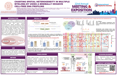
CNA profiles from PB and BM were filtered for tumor fraction ≥3% and a mean absolute deviation (MAD) < 0.20, enabling the selection of high-quality, low-noise profiles for accurate analysis. Comparison of the two compartments revealed a median concordance of 83.3% (range: 26.6–100%), resulting in the identification of two patient groups: those with predominantly unmatched profiles (33/62; 53%) and those with predominantly matched profiles (29/62; 47%). Concordance between cfDNA and gDNA profiles was directly correlated with tumor fraction (median ctDNA in unmatched vs. matched groups: vs. 3.99% vs. 12.06%; r = 0.60; p = 3.63×10⁻⁷).
Previously, we found that high ctDNA levels were linked to diffuse disease, including EM, PS, and FL lesions. In this study, patients with spatially distinct lesions—especially PS and FL with Deauville score 5—showed greater discordance between cfDNA and BM profiles (PS: 67 vs. 85%, p = 0.0268; FL: 72 vs. 90%, p = 0.0210). PET-positive patients (EM, PS, or FL with DS = 5) had larger, more active lesions, with higher SUV values correlating with greater BM–PB genomic mismatch (p = 0.04).
Conclusions:
cfDNA profiling provides a non-invasive way to capture multiple myeloma's genomic complexity and may reflect spatial heterogeneity. Lower concordance with bone marrow profiles is associated with high tumor burden and active lesions. cfDNA can complement biopsies and aid personalized monitoring and treatment.
Acknowledgements: AIRC – Associazione Italiana Ricerca sul Cancro, AIL – Associazione Italiana Leucemia, Linfomi e Mieloma, Bologna.