
Myeloma Genomics and Microenvironment and immune profiling
Category: Myeloma Genomics and Microenvironment and immune profiling
The PBX1 protein is substantially overexpressed in patients with multiple myeloma exhibiting chromosomal 1q gain or amplification, and it is associated with unfavorable prognosis
Pre-B cell leukemia factor 1 (PBX1) gene, located at chromosome 1q23.3, is a transcription factor that modulates essential oncogenic pathways in multiple myeloma (MM), serves as a negative prognostic factor, and may function as a therapeutic target.
Methods:
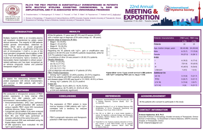
We conducted a retrospective analysis of PBX1 protein expression in bone marrow (BM) biopsies of MM patients with known chromosome 1 abnormalities (1q21+). The immunohistochemical (IH) staining was performed on 3 μm thick sections from paraffin embedded BM biopsies, utilizing a specific PBX1 clone 4A2 monoclonal antibody (Abnova, Taipei, Taiwan, 1mg/mL, M01). In addition, cytogenetic abnormalities (CA) were assessed using fluorescence in situ hybridization (FISH) on CD138-positive BM plasma cells. FISH analysis and BM IH staining were performed on samples obtained at the same time point. Overall survival (OS) was determined with the Kaplan-Meier method. Fifty-four BM specimens from MM patients were analyzed, comprising 45 individuals with 1q21+ and 9 patients without 1q21+.
Results: Of the 54 patients, 31 were men (57.4%) and 23 women (42.6%) with a median age at diagnosis of 64 years (range, 42 - 86 years). Disease stage at diagnosis defined by R-ISS was I in 10 (18.5%) patients, II in 23 (42.6%) patients, III in 19 (35.2%) patients, while for 2 (3.7%) patients it was unknown. Regarding the 45 patients with 1q21+, gain or amplification was present in 29 (64.4%) and 13 (28,9%) patients respectively, while for 3 (6.7%) patients it was unknown. Co-existence of other CA was present in 28 (62.2%) patients. Detection of TP53 was found in 10 (22.2%) patients while t(4;14), t(14;16) and t(11;14) were found in 8 (17.7%), 5 (11.1%) and 5 (11.1%) patients respectively. Lactate dehydrogenase (LDH) at diagnosis was elevated in 17 patients (37.8%). PBX1 protein was expressed in 22 (49%) patients with 1q21+, whereas 23 (51%) were negative. Half of the patients with PBX1 positivity had additional CA and disease stage III R-ISS was found in 10 (10/22, 45.5%) of them. Of the 9 patients lacking 1q21+, PBX1 protein was expressed in 2 (2/9, 22.2%) patients. The estimated 5-year OS for the patients who expressed PBX1 was 34.4% (95% CI: 16.2%-72.8%) versus 48.7% (95% CI: 25.8%-91.9%) for PBX1 negative patients (p 0.54).
Conclusions: The PBX1 protein expression in BM biopsies of MM patients with 1q21+ appears to correlate with a poor trend in overall survival. Further research of the effect of PBX1 expression in the clinical outcome of patients with MM is needed.