
Myeloma Genomics and Microenvironment and immune profiling
Category: Myeloma Genomics and Microenvironment and immune profiling
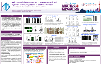
A vicious cycle between sensory nerve outgrowth and myeloma tumor progression in the bone marrow

Motosumi Nakagawa, DDS
PhD student
Tokushima University
Bone pain is a common complication in patients with multiple myeloma (MM). Sensory nerve (SN) fibers have been reported to increase and intermingle with MM cells in bone marrow in MM models. However, interactions between MM cells and SNs have not been studied. In the present study, we aimed to explore SN spreading in the bone marrow and clarify the underlying mechanisms of SN neurite outgrowth in MM and the role of SNs in MM progression.
Methods:
Mouse and human MM cell lines were inoculated into the bone marrow of femurs or tibiae in SCID mice to generate in vivo MM models. Femurs or tibiae were resected, and immunostained after optical clearing with CUBIC method. CGRP+ SNs in bone were then visualized by a light sheet microscope. Neurite extension was analyzed with an axon investigation system. MM cell-specific secreted proteins were selected by a comprehensive proteome analysis for culture supernatants.
Results:
The distribution and density of CGRP+ SN fibers were substantially increased in the bone marrow of femurs or tibiae with MM lesions. MM cell supernatants enhanced neurite sprouting and outgrowth of SNs. Among MM cell-specific secreted proteins, neogenin1 (NEO1) stimulated neurite sprouting and outgrowth SNs derived from DRG cells as well as SN cell lines. NEO1 was constitutively overexpressed in MM cells in all 30 patients’ bone marrow samples tested. NEO1-knockout (KO) RPMI8226 cells mitigated their neurite outgrowth in vitro and reduced the density of SN fibers in MM bone marrow lesions in vivo, suggesting soluble NEO1 as an MM cell-derived SN elongation factor. Membrane-bound NEO1 is a receptor for repulsive guidance molecule-a (RGM-a) which is abundantly contained in sera. rRGM-a inhibited SN neurite outgrowth and acutely retracted elongated neurites in the presence of NGF. However, addition of rNEO1 almost completely abolished the RGM-a’s action. Indeed, rNEO1 and rRGMa formed a complex in a liquid phase as judged by immunoprecipitation. Therefore, soluble NEO1 from MM cells is suggested to induce neurite outgrowth at least in part through acting as a decoy receptor to inhibit RGMa. Intriguingly, the proliferation of MM cells was enhanced in cocultures with SNs. Extracellular vesicles (EVs) isolated from SN supernatants showed potent MM cell proliferation activity. In contrast, pharmacological denervation of SNs by repeated administration of high-dose capsaicin substantially suppressed MM growth in 5TGM1 and JJN3 MM-bearing mouse models. Moreover, NEO1-KO cell growth was attenuated in in vivo models, although rNEO1 did not affect MM growth in vitro.
Conclusions:
MM cells induce neurite hyperplasia in the bone marrow which may increase SN sensitivity to pain and promote MM progression, forming a vicious cycle between SN hyperplasia and MM progression. SNs are implicated as a new niche component in MM, as there has been no previous concept of MM progression being controlled by the peripheral nervous system.