
MRD and Biomarkers
Category: MRD and Biomarkers
3D telomere profiling of MRD in liquid biopsy as a predictive marker of disease stability or progression

Yulia Shifrin, PhD
Laboratory Director (ISO 15189)
Tel Genomics Holdings corp.
Post-treatment, minimal residual disease (MRD) is an FDA-approved endpoint in multiple myeloma (MM) patients. The ability to accurately assess the biological behavior of MRD is limited, as current technologies primarily focus on detecting and quantifying unique DNA sequences or surface antigens of tumor cells isolated from bone marrow specimens, and are unable to predict the biological patterns of these tumor cells.
The evaluation of circulating tumor cells (CTCs) from peripheral blood, rather than bone marrow, offers a promising, less invasive biomarker for MM that allows for continuous monitoring of patients. However, due to the heterogeneity of MM, CTC enumeration alone cannot give a precise indication of MRD stability/progression. We recently demonstrated that the 3-dimensional (3D) profiles of telomeres, a known marker of cancer-associated genome instability, can accurately predict disease progression in patients with smoldering multiple myeloma (PMID: 38747543).
Our current study describes a new workflow for MRD evaluation that combines the enumeration and immunophenotyping of individual MM CTCs with 3D telomere profiling to characterize the residual MM cells or clones, determine MRD negativity or positivity, predict disease progression, and enable continuous non-invasive follow-up.
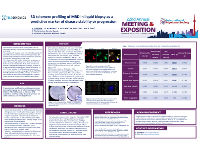
Methods: We report a technique for isolating intact circulating myeloma plasma cells from the peripheral blood of MM patients at the point of diagnosis, post-induction, and 3 months post-transplant/treatment, with subsequent enumeration and immunophenotyping using CD56 and CD138 markers combined with 3D telomere profiling using the TeloView software platform
Results:
Enumeration of MM CTCs demonstrated a high number of detectable myeloma plasma cells in the peripheral blood samples of MM patients at the point of diagnosis. In contrast, the number of detectable CTCs was dramatically lower post-induction and further decreased 3 months post-transplant/treatment. 3D telomere analysis of the isolated CTCs demonstrated 3D telomere profiles characteristic of MM (PMI D: 33921898, 24466378). The captured CTCs were confirmed through pathology review and immunophenotyping.
Conclusions:
The proposed unique workflow integrates the isolation of circulating tumor cells (CTC) from peripheral blood with quantitative enumeration, detailed immunophenotypic characterization, and high-resolution 3D telomere architecture profiling. This allows for longitudinal and minimally invasive monitoring of MRD in multiple myeloma patients from the time of treatment. Unlike conventional approaches, this platform yields functionally and biologically actionable data on CTCs, providing insights into disease stability or progression beyond simple enumeration, while avoiding the need for repeated bone marrow biopsies.